Scintigraphy is the method by which an image, or a series of images, of the distribution of a radionuclide in tissues, organs or apparatuses of the body can be obtained using a Gamma Scintillation Camera. This method is suitable for studying very different body districts and pathologies. For example, we have the use of bone scintigraphy for cancer staging or myocardial scintigraphy for the study of cardiovascular diseases.
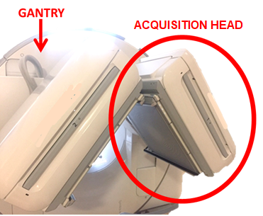
Imaging takes place after the injection of a specific radiopharmaceutical to highlight functional characteristics of the organs and systems under investigation. The images of interest are processed from the measurement of radiation leaving the patient’s body, which is recorded by a special instrumentation called Gamma Camera. The Gamma Camera consists of one or more acquisition heads containing a scintillator crystal and numerous photomultiplier tubes mounted on a gantry so that they can rotate and be positioned around the patient.
The scintillator crystal is used to convert the radiation emitted by the patient into light, which is then converted by the photomultiplier tubes into an electrical signal. The signal is then sent to a computer that uses it for image reconstruction. In new generation devices we also find other elements, the way radiation is converted into a signal may change but the process does not change. Each head can generate a 2D image but, using proper acquisition methods, it is possible to obtain 3D images and dynamic images, i.e., actual movies of how the radiopharmaceutical is distributed inside the patient. The most widely used radionuclide is technetium-99 metastable (Tc99m), which, due to the gamma ray emitted during the decay, and with a half-life of about 6 hours, is the widely used for numerous radiopharmaceuticals:
- for oncological patients, it is used methylene-diphosphonate linked with Tc99m(99mTc-MDP) for bone scintigraphies and Tc99m-linked mercapto-acetyl-triglycine (99mTc-MAG3) for renal scintigraphies
- for heart disease, it is used the molecule sestamibi or tetrofosmin linked with Tc99m.
The radiopharmaceutical is normally injected intravenously into the patient, and depending on the type of examination, Gamma Camera acquisition can occur immediately after injection or even several hours later. Acquisition with the Gamma Camera generally takes from a few minutes to a half an hour.

