PET is a diagnostic imaging examination that uses ionising radiation: it is performed in the Nuclear Medicine department and it is non-invasive and not painful. The term PET (Positron Emission Tomography) is used to refer both to the examination itself and the machine used to perform it.
What is this examination for?
PET examinations are used to obtain functional images of the body: this means that the information obtained is not about the ‘shape’ of organs and body structures (i.e. anatomy), but about their metabolism and ‘functioning’ (i.e. physiology). For this reason, it is a useful examination in the field of oncology (but not only) because it allows to identify the location of any malignant tumour formations in the body.
How does the PET examination work?
PET exploits the decay of special radiopharmaceuticals, composed of so-called ‘positron-emitting radionuclides’ bound to specific molecules.
The radiopharmaceutical is injected into the patient intravenously and is thus distributed throughout the body. Thanks to the ‘tracer’ molecule, however, the radiopharmaceutical accumulates extremely selectively in certain parts of the body, such as in the bladder and at tumour sites.
After a characteristic time, the radiopharmaceutical, as it decays, emits ‘radiation’ called positrons. A positron is a particle like the electron, but with an opposite electric charge: when a positron meets an electron, photons are produced, and the PET scanner is able to record these photons and transform them into images.
The most widely used radiopharmaceutical in PET is Fluorine-18 labelled with glucose (called 18F-FDG fluorodeoxyglucose). Metabolically more active cells need sugars and therefore accumulate more 18F-FDG than less active cells. Cancer cells generally show increased glucose metabolism so the radiopharmaceutical concentrates right inside them and gets trapped: this is the trick used to make the images! Moreover, the half-life of 18F-FDG is relatively short, just under 2 hours, which means that a few hours after the examination the amount of radionuclide present in the patient’s body will be negligible.
The radiopharmaceutical is normally injected intravenously into the patient and, depending on the type of examination, PET acquisition will take place immediately after injection or within a few tens of minutes. PET acquisition generally takes between 10 and 40 minutes.
How is the PET scanner composed?
The PET scanner shown in picture has photon detectors mounted in a circular ring around the patient couch. The machine converts the emitted radiation into an electrical signal and then into images. The images obtained are anatomical ‘slices’ of the patient and depict the 3D distribution of radiopharmaceutical uptake.
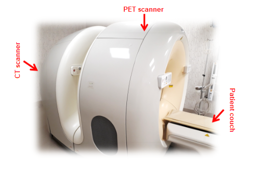
In more recent equipment, the PET scanner is combined with a CT scanner, which is normally used in radiology to acquire morphological images of the human body using X-rays. The fusion of the images produced by these two devices results in PET-CT studies, which are reconstructions of functional and morphological images of the patient.

