
Page 56 - Fisica in Medicina n° 4 - 2017
P. 56
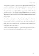
volume structures (RS) and the CT images. Then, a cine acquisition in air with the EPID of the beams of this plan is performed. At the SCH these acquisitions are scheduled in the RT Chart platform (Varian Medical Systems, Palo Alto, USA), usually in the same session of the acquisition of the integrated images to be used for ED. The images are acquired with the EPID at SDD = 150 cm, as shown in Figure 1a. Then, those images are transfered as RI DICOM files using the DICOM export wizard tool in RT Chart. These files are saved in the department network inside a specific folder of the patient. These images are then transformed into RMU maps using the DC task IMAT (ConvertIMATImages), from the main window. The plan, the beam, the deconvolution kernel and the reference 10X10 cm2 field have to be selected accordingly. The reference field is obtained with 200 MU, which needs to be stated in the toolbar. DC performs the procedure described previously to obtain the RMU map, as shown in Figure 1b. Finally, the dose is calculated in the patient’s CT as explained previously and shown in Figure 1c, from which three dimensional dose information can be compared to the TPS calculations.
